The Quadriceps
If you are new to anatomy, the mass of muscle at the front of the thigh was initially thought to be made up of four muscles tied together. And so collectively it’s called the quadriceps. It includes three vastus muscles that work solely on the knee. And it includes the rectus femoris which works on the knee and the hip.
More recently a fifth muscle has been discovered, the tensor vastus intermedius.
In addition, both the vastus lateralis and the vastus medialis have a distinguishable lower portion that can be used to stabilize the knee laterally and medially. These are called the vastus lateralis obliquus and the vastus medialis obliquus.
And so the quadriceps is, quite frequently made up of more than four muscles.
The Vastus muscles
The three usual vastus muscles are called:
- vastus medialis,
- vastus intermedius and
- vastus lateralis.
Their names derive from their position on the front of the thigh.
Vastus medialis is the medial or middle-wards muscle.
It’s the tear drop shaped muscle and the front inner corner of the thigh.
The Vastus lateralis is the lateral or outwards muscle. It forms the bulk of muscle that runs along the outer thigh.
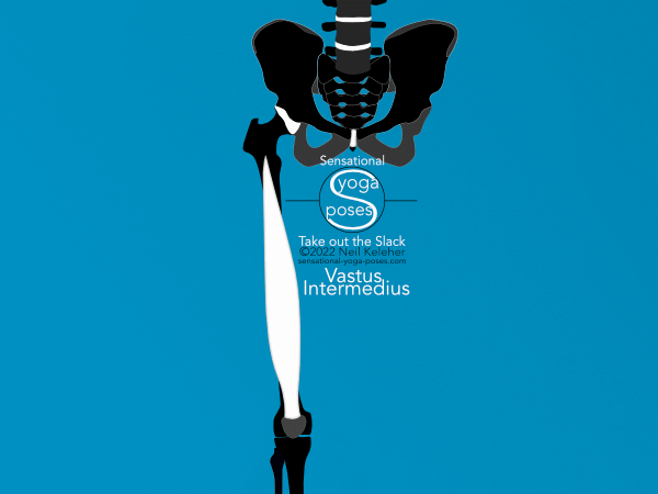
Then there’s the Vastus intermedius. It’s positioned along the front of the thigh. Directly over it is the rectus femoris which also runs down the front of the thigh.
As for the tensor vastus intermedius, it is situated between the vastus intermedius and vastus lateralis. It generally attaches to the top of the patella, towards the inner or medial edge.
Driving Knee Extension or Preventing Knee Flexion
In general, the vastus muscles along with the rectus femoris act to straighten the knee, which in technical terminology is termed extension.
They also resist bending of the knee, which can be thought of as flexion.
- So if you are bending your knees to squat, the quadriceps can help to control the bend of the knee by gradually lengthening so that you can squat down slowly.
- Straightening your knees to stand back up, the quadriceps can gradually shorten.
- If you hold a squat position, say mid-way between fully bent and fully straight, your quads will stay active and remin the same length.
The Lateral and Medial Vastus Muscles help control knee rotation
While it is generally understood that the knees can bend and straighten, it’s important to realize that they also rotate. It’s part of the reason we have three vastus muscles on each leg. The lateral and medial heads help to control the degree of knee rotation.
They also help to keep the knee cap tracking correctly relative to the femur.
Note that knee rotation, that is, rotation of the shin (or lower leg bones) relative to the thigh bone, generally only occurs when the knee is bent. However, even when the knee is straight, the muscles that control knee rotation can still be active. They help to stabilize the knee joint whether it is bent or straight.
Apart from controlling knee rotation, another way to think of these muscles is that they allow the knee joint to transmit rotational forces from the foot and the hip and vice versa without being torn apart in the process.
Knee Pain and the Vastus Medialis Obliquus
With regards to knee stability and knee pain, one muscle that gets a lot of attention is the vastus medialis, the inner vastus muscle.
It’s been noted in some studies that knee pain seems to occur in knees where the vastus medialis fails to activate.
A portion of the muscle that is particularly associated with knee pain is the vastus medialis obliquus. This is the lowest portion of the muscle.
I’ll suggest here that vastus medialis (and vastus medialis obliquus) activation is helpful in dealing with knee pain. Part of the key to getting the bottom portion of the vastus medialis muscle to activate is to first activate the adductor magnus long head muscle.
Why? Because the fibers of the vastus medialis obliquus attach to the tendon of the adductor magnus long head muscle.
If that muscle is active, then its tendon is tensioned and that means the obliquus fibers have a stable or fixed end point from which to activate.
How the Vastus medialis attaches to the Quadriceps Tendon
The quadriceps tendon is the tendon that attaches the quadriceps to the knee cap. The superficial layer is provided by the rectus femoris tendon. The layer below that comes from the vastus lateral aponeurosis. The three layers below that come from the lateral and medial aponeurosis of the vastus intermedius.
The vastus medialis isn't actually a part of the architecture of the quadriceps tendon. Instead it's fibers attach to the aponeurosis of the rectus femoris and vastus intermedius (and to the medial border of the knee cap).
So the vastus lateralis and intermedius generate a lateral and upwards pull on the knee cap. From there the vastus medialis can be used to provide the required degree of medial pull.
To understand what this can mean, imagine that you are using a crane to lift a grand piano to your new apartment on the 4th floor.
There is a main cable that does the lifting, but there are also smaller cables that your team can pull on to orient the piano and to pull it one way or the other. Note that to prevent the piano from swinging or turning, particularly if it's a windy day or the crane operator is less than smooth, it helps if they keep tension on their cables.
The vastus medialis is the equivalent of one of these auxillary cables. It is used to correct the position and alignment of the knee cap, and to help maintain it.
While it doesn’t do the main lifting, it’s still important for adjusting and maintaining the position (or tracking) of the knee cap.
Saying that the vastus medialis is not part of the architecture of the quadriceps tendon may or may not be important.
In terms of sequencing muscle activation of the adductor magnus long head and the various vastus muscles:
- We could activate the adductor magnus long head first so that the vastus medialis is ready to activate.
- Then we can activate the vastus intermedius and lateralis.
- Then the vastus medialis can activate if required.
More on the Aponeurosis of the Vastus Medialis
This is from Clinical Anatomy of the Quadriceps Femoris and Extensor Apparatus of the Knee:
Aside from attaching to the quadriceps tendon, the aponeurosis of the vastus medialis attaches to the medial edge of the patella. These fibers extend further downwards then any other part of the quadriceps tendon. Some of these fibers reinforce the joint capsule as part of the medial patellar retinaculum.
Layers of the Quadriceps Tendon
It’s easy to think, because it is so obvious, that the rectus femoris creates the main upwards pull on the knee cap. In the same study mentioned previously, it was shown that the vastus intermedius contributes three layers to the quadriceps tendon: a deep layer, and two intermediate layers.
The vastus lateralis contributes an additional layer above those. Then the rectus femoris contributes the most superficial layer.
Even though the rectus femoris is perhaps the most obvious contributor to the quadriceps tendon based on external appearances, it is the most superficial contributor. This means that it is not the primary force generator for the quadriceps tendon. It is thus not the main entity respoinsible for generating an upwards pull on the knee cap.
That would be the vastus intermedius (and the vastus lateralis).
Contrasting rectus femoris and vastus intermedius function
One point about the rectus femoris is that it works on both the knee and the hip. And so one problem with viewing the rectus femoris as a primary force generator for the knee cap is that it is strongly affected by the position of the hip joint.
If the hip joint is bent forwards or flexed, this reduces the length of the rectus femoris meaning that it may not be able to activate effectively.
And so the vastus intermedius, buried beneath the rectus femoris, may be the primary force generator for the knee cap.
Now what good is this understanding?
Comparing the medial head of the triceps and the vastus intermedius
For myself, I recently had elbow bursitis. It wasn’t caused by infection. Rather it was caused by incorrect exercise technique.
Actually, it wasn’t caused by incorrect exercise technique, it was caused by muscle imbalance.
I only got it on one side. I got it on the side that I’ve known has had problems.
Doing some research I found out that the attachment of the medial head of the triceps is deep to the superficial triceps tendon. And the medial biceps on my arm that had the bursitis wasn’t functioning.
So I've been spending a lot of time trying to get my medial triceps to activate.
In the process I wondered if the knee joint didn’t have a similiar structure. That’s when I found the study on quadriceps tendon structure and found out that the vastus intermedius, rough equivalent of the triceps medial head, has a deeper attachment to the knee cap than the other quadriceps muscles.
Rather than just memorizing names of muscles and their origin and insertion, I study anatomy so that I can draw it, if required, and more importantly so that I can attempt to feel it and control it. This is generally to try to fix problems, but also to try to improve function. A general side effect is that it tends to improve my understanding of how muscles in general work, and how they work together in the context of my own body. And so after reading about the vastus intermedius and how it provides the deepest layer of the quadriceps tendon, I looked at how to activate it. I also tried to notice if it helped my knees in any way.
How to activate the vastus intermedius muscle
So how do you voluntarily activate the vastus intermedius?
One trick to activating the vastus intermedius is to create an upwards pull on the back edge of the top of the knee cap.
Depending on your position, standing or sitting, knees straight or bent, you may find it helpful to direct the pull on the knee cap towards the front surface of the thigh.
Or you could try "sucking" the front of your thigh forwards.
Try to generate the "sucking" action along the entire front surface of the femur, from the knee cap up to its neck.
Do this in combination with pulling on the back edge of your knee cap.
To try to isolate it, you can try to keep your hip joint relaxed while activating your vastus intermedius. (Try this while sitting.)
Generally with muscle control, if you are specific with your point of pull, you can target particular muscles and activate them.
So for example, with the triceps medial head, one possible means of activating it is generating a pull on the inner corner of the elbow. Another means is to generate a pull closer to the joint (closer to the back surface of the upper arm bone).
Later on, I found out that activating pronator teres first was a sure fire way, at least for me, for activating the medial head of the triceps.
Activating the Vastus muscles in general
Because the vastus muscles attach to the top and side edges of the knee cap, you could aim at a fuller quadriceps activation by generating a pull on the top of the knee cap and its outer edge to activate the vastus intermedius and vastus lateralis. From there, you could generate a pull on the inner edge to activate the vastus medialis.
You could then work at fine tuning, or adjusting, the amount of vastus medials activation. Likewise, the amount of vastus intermedius and vastus lateralis activation.
Vastus intermedius activation, Improving knee function, reducing knee pain
As hinted at previously, one of the main reasons that I was interested in learning about the vastus intermedius is that I've had knee pain.
I've gradually fixed various knee pain problems through learning to control various muscles.
While I've mostly fixed my knee problems, I still have niggling problems on one side. And so after my experience with elbow bursitis I was interested to see if a similiar activation with respect to the knee would improve my knee function and lessen the incidence of knee discomfort.
Note that learning to activate the vastus intermedius (or any muscle for that part) in isolation is generally the first step. From there the challenge includes activating it under different conditions.
he challenge the is integrating (or re-integrating) i.e. activating with other muscles.
Activation protocols for the legs
As mentioned, the knee is part of a force delivery system that includes the hip, the joints of the foot and ankle and possibly the SI joint. What can be a good practice when working on any part of the leg is to practice activating that part as part of a sequence of activations that involve the other joints of the lower limb.
Generally when working with the legs I like to sequence activations working from the hip (or from the SI joint) down, or from the foot up.
With the hip joint, one important sub system is the hip joint suspension system. When working from the hip to the foot, I generally activate the hip suspension system first.
Secondary to this is activating the adductor magnus long head so that the vastus medialis obliquus is ready for action.
From there it may be wise to activate the vastus intermedius. And then from there, if necessary the foot. (My current protocol for foot activation involves activating either the inner foot or the outer foot-and-heel, or both. )
It may be that the adductor magnus is itself part of a subsystem that includes the pes anserinus muscles and may also include the popliteus and biceps femoris short head. And so while I tend to think in terms of the adductor magnus long head, it may be that I'm activating it and the sartorius and other pes ansernus muscles.
Working from the foot upwards, you could reverse these steps. Whether working from the hip downwards or the foot upwards, an important point is to adjust or fine tune your muscle activations as well as your positioning and alignment.
With respect to alignment, the idea is to adjust not based on external appearances, but instead based on feel. In this case feel refers to the forces generated by muscle activation and transmitted by connective tissue. It also includes the forces at points of contact.
The important point in all cases is being able to feel what you are activating.
What you are trying to feel are the forces generated by muscle activation and transmitted via connective tissues.
Extending this field of sensitivity, you can further feel skin contact and changes in pressure where ever parts of your body come in contact with itself or anything (or anyone) else.
If you can feel each activation, you can then adjust or fine tune it. In addition, as you get more practice, and more experience, you may find that you can play with the sequence of activations. You’ll then be able to self-determine whether a particular sequence of activations is appropriate simply based on the feel.
References
Clinical Anatomy of the Quadriceps Femoris and Extensor Apparatus of the Knee
New insight in the architecture of the quadriceps tendon
Published: 2022 04 24


