Shin rolling (relative to the foot) and the tibialis posterior
One action you can do while standing is rolling the shins inwards and outwards. Assuming the inner edge of the forefoot is kept in contact with the ground:
- Rolling the shins out accentuates the inner arch of the foot.
- Rolling the shins out flattens the inner arch of the foot.
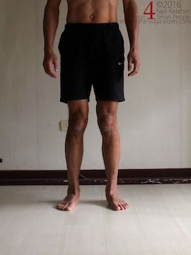
Shins rotated inwards
(Tibialis Posterior possibly relaxed)
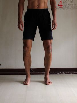
Shins rotated outwards
(Tibialis posterior activated)
Shin rolling can be caused by the muscles that act on the IT Band and the muscles that form the pes anserinus (collectively, the long thigh muscles), particularly if you focus on turning the upper part of the tibia.
However, if the focus is on using the feet to roll the shins, then one of the muscles involved in rolling the shins externally is the tibialis posterior.
One sign of an activated Tibialis Posterior
One sign of an active tibialis posterior is a feeling of muscular tension running up the back of the lower leg bones.
This sensation is "closer to the bone" than the sensation generated when the larger calf muscles (soleus and/or gastrocnemius) activate.
Letting Gravity do Some of the Work
While standing, internal shin rotation can be gravity driven. You simply relax the tibialis posterior and let body weight do the work.
Tibialis Posterior Connections
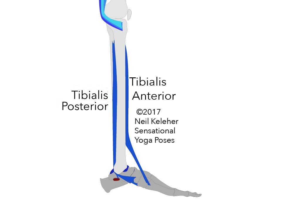
Tibialis Posterior attaches to a large portion of the back surfaces of both the fibula and tibia.
The Tibia is the larger and more substantial of the two lower leg bones. It mates directly with the femur or thigh bone.
The Fibula is a slender bone that runs along the outside of the lower leg. It doesn't bear weight but it as important bone for transmitting tension between the hip and femur and the foot and ankle.
The Lower Tendon of the Tibialis Posterior
The tendon of the tibialis posterior passes down the inside of the ankle, behind the medial malleosus which is the bump at the inside of the ankle which is actually part of the tibia.
The tendon of the tibialis posterior has several branches and thus, several points of attachment.
Because it passes behind the medial malleosus, when activated while standing, the tibialis posterior creates a forward pushing force on the inside of the tibia. Anchored by its attachment to the bones of the foot, this muscle can help to externally rotate the shin or resist internal rotation.
After passing the medial malleosus the tibialis posterior reaches forwards and downwards to attach along the underside of the inner arch of the foot.
Note that while it passes behind the medial malleosus, the tendon of the tibialis posterior passes in front of the sustentaculum tali, a projection on the inside of the calcaneus.
The calcaneus could be thought of as "The Heel Bone."
The talus could be thought of as the "ankle bone". It sits on top of the calcaneus and it's upper most surface supports the bottom of the tibia.
Attachments of the Tibialis Posterior
The tendon at the lower end of the tibialis posterior has several branches.
Sustentaculum Tali
One branch of the tibialis posterior tendon reaches back to attach to the sustentaculum tali as it passes it.
When tibialis posterior is active, this branch can create an upwards pull on the inside of the calcaneus (via the sustentaculum tali).
While standing, this would create an upwards lift on the inside edge of the heel.
Tension on this branch of the tibialis posterior could also create a forwards pull on the inside of the heel. The combined action of pulling the inside of the heel forwards and upwards can assist in the rotating the shin externally relative to the foot when the foot is bearing weight.
Tension on this branch of the tibialis posterior tendon could also be sufficient to prevent the inner heel from sinking or collapsing (in turn helping to support the back end of the inner arch of the foot). This would also help prevent internal rotation of the shin.
Because this part of the tibialis posterior tendon reaches back, it may serve as an anchor for the forward reaching branches of the tendon.
Navicular
The navicular bone butts up against the front surface of the talus bone. The three inner toes (the three largest) all radiate forwards from the navicular bone.
One branch of the tibialis posterior tendon attaches to the medial surface of this bone at the medial tubercle. Tension on this branch of the tendon creates a rearwards pull on the inside edge of the navicular. This tension could help to accentuate the inner arch of the foot and prevent it from flattening.
Medial Cuneiform
Jutting forwards from the navicular are the three cuneiform bones which are positioned next to each other. The inner most of these bones is called the medial cuneiform and is the first distinct bone of the big toe.
Another branch of the tibialis posterior tendon attaches to this bone. Tension on this branch further helps to accentuate the inner arch of the foot or prevent it from flattening.
The Cuboid and More
Attached to the front surface of the calcaneus is the cuboid bone.
This bone sits next to the navicular and the outermost of the three cuneiform bones.
Another set of branches of the tibialis posterior tendon attach to the underside of the two outer most cuneiform bones, the underside of the cuboid and in addition, the underside of metatarsals 2, 3 and 4.
These bones attach to the fronts of the innermost cuneiform bones and the cuboid.
When tension is added to these branches, the inner arch of the foot is further accentuated and supported.
Tibialis Posterior and Foot Posture
While standing the tibialis posterior helps to lift the inner arch of the foot and rotate the shin externally relative to the foot.

Shins rotated inwards
(Tibialis Posterior possibly relaxed)

Shins rotated outwards
(Tibialis posterior activated)
This action can be useful when balancing on one foot. It can also be useful when squatting or doing poses like chair pose and other bent knee standing poses like side angle. And it can be handy when doing standing hamstring stretching poses like pyramid pose, revolving triangle and triangle pose.
The tibialis posterior can be active when the inner arch is flat, helping to stabilize the foot in the "flattened" position.
As well as causing external rotation of the shin this muscle can help resist internal rotation of the shin.
Tibialis Posterior Activation in Non-Weight Bearing Foot Positions.
While seated, say with legs straight out in front, tibialis posterior could be used to turn the soles of the feet inwards.
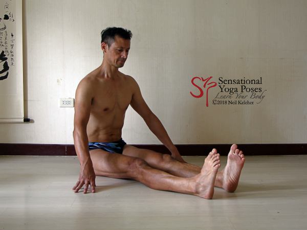
Staff pose: Feet turned inwards with the aid of Tibialis Posterior
In a position like bound angle pose, with knees bent and feet together just in front of the groin, the tibialis posterior muscle can activate to turn the soles of the feet upwards so that the inner edges of both feet move away from each other.
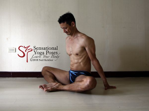
Bound angle pose with soles of feet turned upwards with the aid of tibialis posterior.
It can perform the same foot forming action on the front foot in pigeon pose (particularly in the variation where the front leg hip is on the floor.)
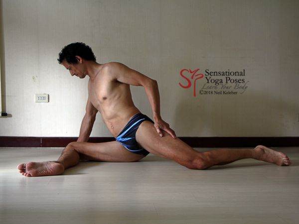
Pigeon pose with front hip on the floor and shin parallel to the front, sole of front foot partially turned upwards with aid of tibialis posterior.
In these non-weight bearing foot positions, tibialis posterior activation can create a strong sensation along the inside arch of the foot.
Published: 2020 08 05
Updated: 2022 05 13
 Shins rotated inwards
 Shins rotated outwards


