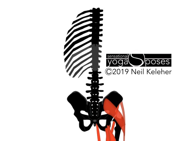
The part of the spine that joints the two halves of the pelvis together is called the sacrum. It looks like a downward pointing arrow with the tip (called the tailbone or coccyx) just behind the anus. The connections between the sacrum and the two hip bones are called the SI joints.
As well as connecting at the SI joints, the two halves of the hip bone also connect at the pubic synthesis or pubic bone. These joints allow the hip bones and sacrum to move relative to each other.
Understanding the muscles that can affect the SI joints and thus the spine
An important part of spinal anatomy can be understanding how various muscles can affect the hip bones, sacrum and thus the SI joints. These muscles include the pelvic floor muscles, the transverse abdominis, the lumbar multifidus and the spinal erectors. The long hip muscles are muscles that attach between the lower leg bones and the corner points of the hip bone. And so muscle activation (or lack thereof) in the feet and ankles can have an affect on the si joints and thus the spine.
The lumbar spine connects the pelvis to the ribcage and is made up of five vertebrae. These vertebrae are quite large and contrary to the usual notion that they are large to support the weight of the upper torso, I'd suggest that they are large so as to be able to transfer forces between ribcage and pelvis and vice versa.
If dealing with low back pain it can be helpful to define the lower back as including the lumbar spine, the sacrum, the si joints, the lower part of the ribcage and thoracic spine (the thoracolumbar junction).
One structure that includes all of this is the thoracolumbar fascia.
One reason for including the sacrum and SI joints in the lower back is that imbalances between the SI joints may cause low back pain. Note this imbalance may actually come from the hip joints, or even from the lower leg bones via the previously mentioned long hip muscles.
Another possible source for low back pain may be imbalance or poor muscle function at the thoracolumbar junction. Basically this is where the lumbar spine meets the ribcage. Muscles that overlap this junction include the latissimus dorsai, serratus posterior inferior and the transverse abdominis.
Note that another structure that can have a possible affect on the low back via the SI joints is the sacrotuberous ligament.
The psoas attaches the femur to the lumbar vertebrae. When active it creates a forwards and downwards pull on the lumbar vertebrae.
The psoas may become tight through over use. If only one side becomes tight this can cause an unbalanced pull on the lumbar spine which overtime may lead to low back pain.
While doing various psoas stretches and or doing a psoas release may help in the short term, the better solution would be to look at the cause of hip imbalance.
When dealing with problems like low back pain it can be helpful to view the sacrum as an extension of the lumbar spine.
If you practice making your spine feel long, you can include the sacrum as you work at making your spine feel long.
If you can feel your sacrum via the muscles that attach to it, (multifidus for example) it can then be relatively easy to feel your SI joints.
Because the spine tends to have curves, if you flatten or straighten these curves, you can make your spine longer.
One way to practice this is with some simple improve your posture exercises.
The actual act of straightening the spine uses various muscles that act between the vertebrae. These include the spinal erectors as well as smaller muscles.
When using the smaller spinal muscles, the muscle activation sensation they create is rather small and so what you can feel or "sense" instead is connective tissue tension as it is lengthened by the action of these muscles.
The key to activating these smaller muscles is to focus on feeling your entire spine.
So that you can work towards feeling your entire spine, practice feeling the intervertebral joints of your spinal column individually.
Another way to learn to feel your spine it so practice moving it. For more on this read feel your spine.
For more on feeling your spine check out feeling your spinal column
Note another way to feel your spine is to practice bending it backwards in a pose like cobra yoga pose. To get even more sensitive, focus on bending it backwards one vertebrae at a time.
For more on this read cobra yoga pose.
The thoracic spine is the part of the spine to which the ribs attach and it has twelve vertebrae. I like to think in terms of a lower and upper thoracic spine.
The lower part of the thoracic spine is the part whose corresponding ribs form the arch at the front of the ribcage. Like the lumbar spine this part has five vertebrae.
The upper part of the thoracic spine has ribs that attach directly to the sternum. This part, like the cervical spine, has seven vertebrae.
The ribs can be divided into three categories, true, false and floating.
The true ribs are the upper seven pairs of ribs that attach directly to the sternum. The false ribs attach to the sternum via cartelide. The floating ribs are the two lower most pair of ribs whose ends are free.
Taken individually, the ribs can be thought of as levers. The obliques and intercostals as well as muscles like the serratus posterior inferior, can act on the spinal vertebrae via the ribs.
Because of this the intercostals and obliques can be used to twist, side bend, front and back bend the ribs, and via the ribs, thoracic vertebrae relative to each other.
In addition, these muscles can work against each other and other muscles to help stabilize the ribs and thoracic spine.
One important reason for the sternum is that it makes the upper ribcage more stable. It can thus act as a foundation for arm and shoulder muscles that attach to it.
As a comparison, some types of turtles developed shells in particular to enable them to dig more efficiently. The shell provides a foundation for stronger digging actions.
In a similar way, if you look at an ostrich or emu skeleton, they have large sternums so that the muscles that work on the wings have a strong foundation. Similarly, we have a sternum so that we can use our arms effectively and so that we can use them to transmit or deal with forces.
Interestingly the shoulder blade tends to float in the region of the upper part of the ribcage, the part that corresponds to the sternum.
One reason we have a ribcage is not so much for protection, though that may be important. As described in the previous section, the reason we have a ribcage in part is to give muscles that work on the arms a foundation so that they can be used more effectively.
Muscles that can act from a stabilized ribcage to help with scapular stabilization include the serratus anterior and pectoralis minor.
Another muscle that attaches more to the spine than the ribcage but that is also involved in stabilizing the shoulder blade (another word for scapula) is the trapezius muscle.
Note that the upper ribcage does tend to be more rigid. However, through the use of opposing muscles it's also possible to stiffen and this stabilize the lower ribcage.
Note that because the ribcage is a little bit flexible, what this means is that the ribcage can be stabilized in a variety if shapes. This gives us greater flexibility in how we can apply our arms and arm strength.
Another reason for the ribcage is as a anchor or foundation for the respiratory diaphragm.
When the bottom rim of the ribcage is stabilized, it can help to anchor the diaphragm making it easier for this muscle to activate.
The cervical spine joins the ribcage to the head and has seven vertebrae.
The previously mentioned trapezius muscle attaches in part to the cervical spine. As a result, to anchor this muscle so that it can act effectively on the shoulder blade it can help to stiffen the neck or, more elegantly, to make it feel long.
For an example of how to make your neck feel long check out these exercises for fixing forward head posture
As a whole the spinal column has two main functions. One is that it acts as a bony support and connecting device for the head, ribs and pelvis. Second, it serves as an information channel carrying nerves from the brain to the rest of the body.
Passages exist between adjacent vertebrae so that nerves can leave the spine channel and venture to other parts of the body.
The main supporting function of the spinal column is carried out by the bodies of the vertebrae which are connected to each other via intervertebral disks. These disks create space between adjacent vertebrae while at the same time keeping them connected and allowing them to move relative to each other.
The intervertebral discs can also act as shock absorbers.
To the rear of the vertebral bodies a channel is formed for the passage of nerves. To the rear of each vertebrae is a spinous process which point rearwards and two transverse processes which angle outwards and backwards.
The spinous processes can limit backward bending of the vertebrae relative to each other.
The transverse processes can limit side bending.
All of the processes serve as points of muscular attachment and so can be used to exert leverage on each vertebrae. In particular they can be used as points of leverage for back bending, side bending and twisting.
What do vertebral joint facets do?
Each spinal vertebrae has upper and lower joint facets which limit and define the movement possibilities of the vertebrae relative to each other.
For example, the lumbar facet joints tend to allow front, back and side bending but restrict rotation. Be aware, this doesn't mean that the lumbar vertebrae can't rotate relative to each other. It just means that rotation is severely limited. That being said, it's still important to be aware of, especially in regards to feeling and controlling the lumbar spine while doing spinal twisting poses and actions.
Various levels and layers of muscles enable the spine to move in different ways.
Working from smaller to larger, the muscles of the spinal column span can span a single vertebral joint or many. Muscles that joint adjacent transverse processes and adjacent spinous processes can help in side bending that spinal segment or back bending it.
Muscles that jump from transverse process to spinous process can also be used to backbend that spinal segment but in addition can twist it.
Larger muscle groups work similiarly but work across two or more spinal segments.
The spinal erectors tend to be longer muscles that work across the back of the pelvis, ribcage and head as well as across the vertebrae. All together these muscles allow the spine a lot of flexibility in bending and twisting in different ways.
These same muscles can also be used to stabilize the spine.
You can read more about these muscles in the intrinsic back muscles article.
One of the main muscle groups that work on the spine is the spinal erectors.
Because the spine attaches to the pelvis, ribcage and head, muscles that work between these structures can also affect the shape and stability of the spine.
- The abdominal muscles affect the relationship between the ribcage and the pelvis.
- The quadratus lumborum affect the relationship between the ribcage, lumbar vertebrae and the lowest pair of ribs.
- The intercostal muscles affect relationships between adjacent ribs and adjacent thoracic vertebrae.
- Muscle that work between the ribcage and neck and between ribcage and head affect the cervical spine.
- In addition muscles that attach from the shoulders to the neck and skull also affect the cervical spine.
- Finally, hip muscles can affect the spine directly if they attach to the sacrum or indirectly if they attach to the hip bones. Muscles that attach to the hip bones include sigle joint hip muscles like the single joint hip flexors and dual joint hip muscles like the long hip flexor muscles or the even more inclusive long hip muscles. These are muscles that attach to the corner points of the hip bone from the lower leg bones and thus can exert a lot of leverage on the hip bones and potentially the SI joint.
Experiencing your spine while walking a twisting
One way that you could get an experience of your spinal anatomy is while walking with a twist. Read more about that and the related spinal anatomy in walking with a twist.
Published: 2020 08 28
Updated: 2020 10 30


