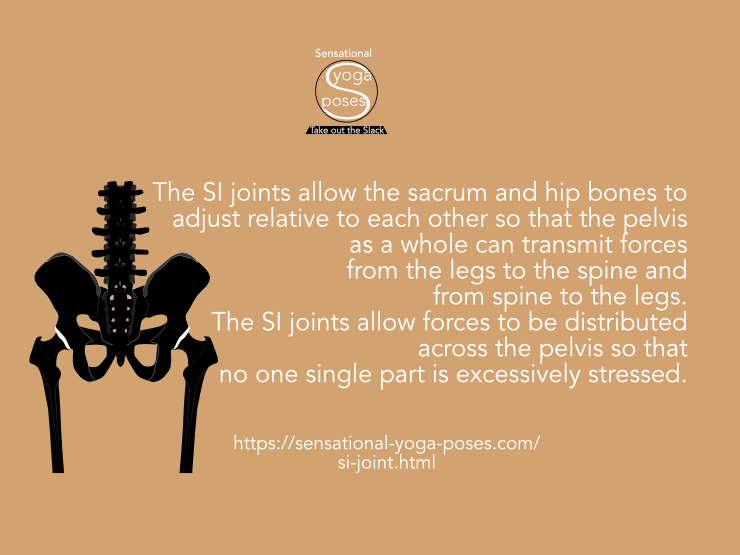
Movements of the Sacrum Relative to the Hip Bones (and Vice Versa)
Two references that are important for describing this movement are the ASICs and Ischial Tuberosities.
Two Sets of Reference Points for the Pelvis
If you put your hands on the front corners of your belly, about an inch below the belly button, you can feel two sharp or pointy bones. If you are overweight, you can approach these bones from the side. These are the fronts of the iliac crest and are referred to as the Anterior Superior Iliac Crest (ASIC).
The Ischial Tuberosities, or sitting bones, are the two bones you can feel when you sit on a hard seat.
Looking at the pelvis from the side, and viewing it as a rough square or trapazoid, if the pubic bone and sacro iliac joints form two opposite corners of this square, then the ASICS and Ischial Tuberosities (or ITs or Sitting bones) form the other set of opposite corners.
Warping the Pelvis
The hip bones connect directly to each other via the pubic synthesis, what we tend to think of as the pubic bone.
Because the two hip bones hinge at the pubic synthesis and at each SI joint the pelvis can flex as follows:
- a nodding movement of the sacrum (nutation) causes the ASICs (and the top of the sacrum) to move inwards and the Sitting bones (and the bottom of the sacrum) to move outwards.
- A backwards nod (counter nutation) of the sacrum causes the ASICs (and the top of the sacrum) to move outward and the sitting bones (and the bottom of the sacrum) to move inwards.
The Muscles that Bend the Pelvis
Nutation/Nodding can be directly caused by the lower third of the transverse abdominus muscle contracting. It may be helped by an activation of the sacral multifidus (a feeling like "flicking" the tailbone rearwards).
Meanwhile, Counter-Nutation/Backward-nodding can be directly caused by the pelvic floor muscles activating.
In my own experience, activation of one set of these muscles automatically causes the other set to activate. You could think of the two (or three) sets of muscles acting against each other to stabilize the SI joints (and pubic synthesis).
In males the movement potential tends to be quite small, in females it tends to be larger. However, the same musculature for moving or stabilizing these three bones exists in both sexes. And even though the movement potential varies across the sexes, the muscular actions that drive these movements can be controlled and felt by both sexes.
(While I would recommend learning to feel these actions, I would not recommend keeping either set of muscles activated on a regular basis. This applies to any set of muscles. Muscles are meant to activate and relax.)
Why Make the Pelvis a Flexible Structure (via the SI joints)? Why?
Why have this movement potential at the SI joints in the first place? Why allow the two halves of the pelvis and the sacrum to move relative to each other?
The body is designed to be maximally strong with minimal use of materials.
This end is achieved at skeletal joints by allowing bones to move relative to each other so that connective tissues (tension elements) can freely redistribute tension.
Maintaining Space in Non-synovial Joints
Non-synovial joints (like the sutures of the skull and also generally termed "fibrous joints") are shaped and tied together by connective tissue in such a way that pressing the two bones towards each other actually causes the tension in the connective tissue to increase in such a way that it resists the bones being pushed together. The more you push the more it pushes back. The same happens if you do the opposite and try to pull the bones apart. Tension increases to resist the pulling.
The SI joints are partial synovial joints.
Maintaining Space in Synovial Joints
Synovial joints use fluid to maintain space and lubricity between the bones they connect.
They work to keep bones from touching via a fluid filled joint capsule whose tension can be varied by muscle controlled ligaments.
In this case the control mechanism is a bit more complex, but the bare-bones description is that muscle tension can be increased to add tension to the joint capsule via ligaments and tendons. That in turn increases fluid pressure which acts to resist the bones being pushed together.
Flexing Your Biceps (And "Stabilizing" your Elbow Joint)
If you've ever "flexed your biceps" like a body builder, you were not only activating your biceps, but also your triceps. The two sets of muscles work against each other to maintain the bend in your elbow. Possibly because you are focused on the biceps (look how big it is), you've never noticed that your triceps was active or that your elbow felt "stable".
Flexing your biceps (and triceps) with the elbow straight, the action of the two sets of muscles would pull the humerus towards the radius and ulna. The same muscle activation that adds tension to the triceps and biceps (and possibly the forearm muscles that act on the elbow) also sends tension to the ligaments (as well as the tendons) and via the ligaments (and possibly the tendons both) adds tension to the joint capsule. This then adds pressure to the synovial fluid inside the elbow joint which resists the bones being pushed against each other.
Maintaining Joint Space, In General
So in the case of pressing two skull bones towards each other, tension within the sutures increases to resist and so maintain space between those bones.
Synovial joints can respond similiarly.
When the very muscles that act on the joint activate to squeeze the bones together, the joint capsule adds pressure to the fluid within it to resist the bones being pushed together.
For a synovial joint, when muscles contract they not only create a gross movement, the tension they create also acts to try to reduce the space between those bones at the joint. And the joint capsule, driven by the same muscle action, responds, by increasing fluid pressure to keep space between the bone.
Maintaining Space Allows Adjustability (and The Equal Redistribution of Tension)
While the bones are kept from touching, tension within the joint capsule can be distributed, in part helped by the pressure of the fluid within the joint capsule. (At least that would be the theory) and in part helped by the fact that the bones aren't touching. Because they aren't touching (or frictioning), they have some adjustability relative to each other again allowing for the joint capsule to redistribute tension within itself.
That redistribution doesn't just happen within the joint capsule. The joint capsule may have ligament and tendon connections. And so tension is redistributed to other parts of the body via these connections also.
The pelvis is broad and that can lead to large force moments
As a whole, the pelvis is the broadest structure in the body.
The legs attach to it and the spine. Because of this broadness, force moments acting on the pelvis can be large.
Adjustability between the hip bones and between each hip bones and the sacrum gives the pelvis enough flexibility to redistribute loads within itself while at the same time functioning effectively as a connecting element between the legs and the spine.
Because of the flexibility given to the pelvis by the SI joints and the pubic synthesis, the pelvis is strong enough to handle these loads and transmit them. The flexibility given to it by the SI joints means that it can make adjustments within itself while transmitting loads so that the tension elements within itself share the load.
Optimal Positioning
Another possible reason for pelvic flexibility is that it could allow for optimal positioning of the hip bones of the pelvis relative to the thighs, particularly in extreme positions like forward or backward bends of the hips. (And even if this movement is slight, it continues to allow effective the redistribution of tension.)
Extending Your Reach, how a mobile shoulder blade can prevents shoulder impingement
As an example of optimal bone positioning, the shoulder blades can more relative to the ribcage depending on how we are using the arms.
To move the arms forwards the shoulder blades more outwards and forwards relative to the ribcage extending the reach and possibilities of the arms in that direction.
To move the arms upwards the shoulder blades rotate upwards.
(If using the shoulder sockets as a reference, this movement could be called supraversion since it causes the "eyes" of the shoulder sockets to look upwards.)
To move the arms back, you can lead by moving the shoulder blades towards each other. (This movement is called retraction, the opposite movement, moving the shoulder blades outwards and forwards is called protraction).
Allowing the shoulder blades to move relative to the ribcage means that the muscles that attach the shoulder blade to the arm bones can be in an optimal position to work no matter what the arms are doing (assuming that everything is working optimally.)
In this case optimal could mean that no muscle is overly long or over short. All muscles are within their optimal working range so that they can activate effectively to help support the arms in whatever they are doing.
Basically it prevents any muscles from "over-reaching" or being too cramped to work and that may help to keep the shoulder joint from being ripped apart. (Or getting back to "tension distribution" it allows the continual redistribution of tension.)
Stop Your hip from Impinging Now!!!
The hip socket is deeper than the shoulder socket and the hips tend to be less mobile than the arms. And so there may be less need for the hip bones to move relative to the spine for optimal muscle length.
However, near the end ranges of movement it might be beneficial to move the hip bones relative to each other, if nothing else to help prevent bones from bumping into each other.
Drawing on the shoulder again as an example, there's a lip of bone above the shoulder socket called the accromion process.
This bone is an extension of the spine of the scapula. It could serve to protect the shoulder joint.
But it also serves as an attachment point for muscles and it is also provides an attachment point for the collar bone.
Whatever the function of this covering of bone, when raising the arm it tends to get in the way. And so to avoid bone to bone contact, this part of the shoulder blade has to be pulled upwards and inwards to give the humerus room to move upwards and inwards.
At the same time the bottom tip of the shoulder blade moves outwards.
But getting the accromion process out of the way of the humerus might only be a side effect of this movement, made obvious when people have problems with the two bones bumping into each other.
Since the accromion process is a point of muscle attachment for the middle trapezius, as well as the delts, this movement of the scapula that is supposed to accompany an arm lift may be more important in preserving muscle function, giving the delts room to contract to help support the arm.
And the same may be true with movements of the pelvis. In extreme movements like the splits, adjustments of the bones of the pelvis may maintain the operability of the muscles that work on the hip joint (as well as preventing the the greater trochanter of the femur from bumping into the side of the pelvis)
And that could be important as a mechanism for protecting the integrity of the hip joint.
Creating a stable foundation for one part of the SI joint or another
Moving the femurs relative to the hip bones, in positions like side to slide splits, or front to back splits, or extreme front bends or back bends at the hips, movements of the hip bones relative to the sacrum can help to prevent bones from impinging. More specifically, it can help prevent the femur from pinching soft tissue between itself and the hip bone.
So that the SI joints can allow these hip bone movements it can help to have a stable reference bone. That could be the femur, or it could be the spine and sacrum. Note that depending on whether the feet are on the ground (and bearing weight) or not, the stable foundation could be the hip bones themselves.
If muscles have a stable foundation from which to act and optimal length, not only can they effectively control bony relationships, they can also effectively control joint capsule tension.
Thus, assuming a stable foundation, movements at the sacrum can be to give muscles optimal length so that not only do they integrate the SI joint but the hip as well.
Published: 2020 08 14


